
“When learning to think about molecules on a 3D atomic level, it is difficult to orient the molecules in the human mind. When using 3D printed models, it trains the brain to orient and visualize the 3D molecules accurately,” explains second-year Biochemistry and Cell Biology graduate student Sarah Alvarado. Last spring, she and other students took Structural Biology (BIOC 482/552), a course taught by Dr. George Phillips. The course was taught on a molecular level forcing students to think about protein-protein and protein-DNA interactions on an atomic level rather than the simplified 2D interactions that are commonly taught in introductory science courses.
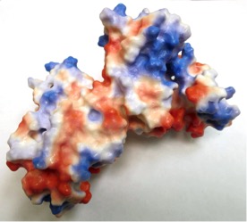
3D Model of VP40 Matrix Protein from Ebola Virus: The red and blue highlights illustrate the electrostatic residues that play a role in the polymerization of this protein during the virus life cycle.
Using Rice’s 3D Printer, she and her classmates printed proteins of their choosing to use as a teaching aid in their end of semester presentations. Alvarado selected two proteins that had been solved, or identified, within the previous calendar year: Ebola virus matrix protein VP40 and a cascade complex that forms as part of the CRISPR/Cas system. She was able to highlight particular aspects of these proteins. For example, the red and blue highlights on the VP40 protein portray the electrostatic forces that are critical in the polymerization of this virus.
On the CRISPR/Cas protein, she decided to print two molecules of the protein and to label each of the eleven corresponding subunits the same color.

3D Model of Cascade Complex Isolated from E.Coli: This 3D model represents two molecules of the CRISPR/Cas protein cascade complex. Each subunit of the complex is indicated by different colors.
In order to do this, Alvarado and other students simply imported the published protein structure sequences from the Research Collaboratory for Structural Bioinformatics Protein Data Bank into Pymol, a program that generated a structure from the sequences. In Pymol, the proteins were edited, rotated in space, and visualized prior to printing. Additionally, Alvarado explains that some molecules needed “struts,” which are small reinforcements to help strengthen the printed protein structures. For example, Alvarado’s CRISPR/Cas coils required these struts. Then, the proteins were sent to the 3D printer. “I was able to gain a better understanding of what is going on at an atomic level and to think of how dynamic the molecules are by holding onto and seeing the proteins,” Alvarado adds. “It is great to have a 3D printer on campus, especially for further understanding concepts instead of simply hearing and trying to visualize them.” Alvarado believes that it would be advantageous for professors to use these 3D models in introductory undergraduate Biology courses “to emphasize how complex these interactions truly are in our bodies” and to provide a better illustration of the 3D nature of atomic interactions. Currently, Alvarado is working to solve protein structures involved in cancer. When she does, she will most definitely return to the 3D printer to illustrate her findings and better portray her work in a more dynamic and visible way.